Diagrams of the Teeth Biology Diagrams Atlas of dental anatomy: fully labeled illustrations of the teeth with dental terminology (orientation, surfaces, cusps, roots numbering systems) and detailed images of each permanent tooth

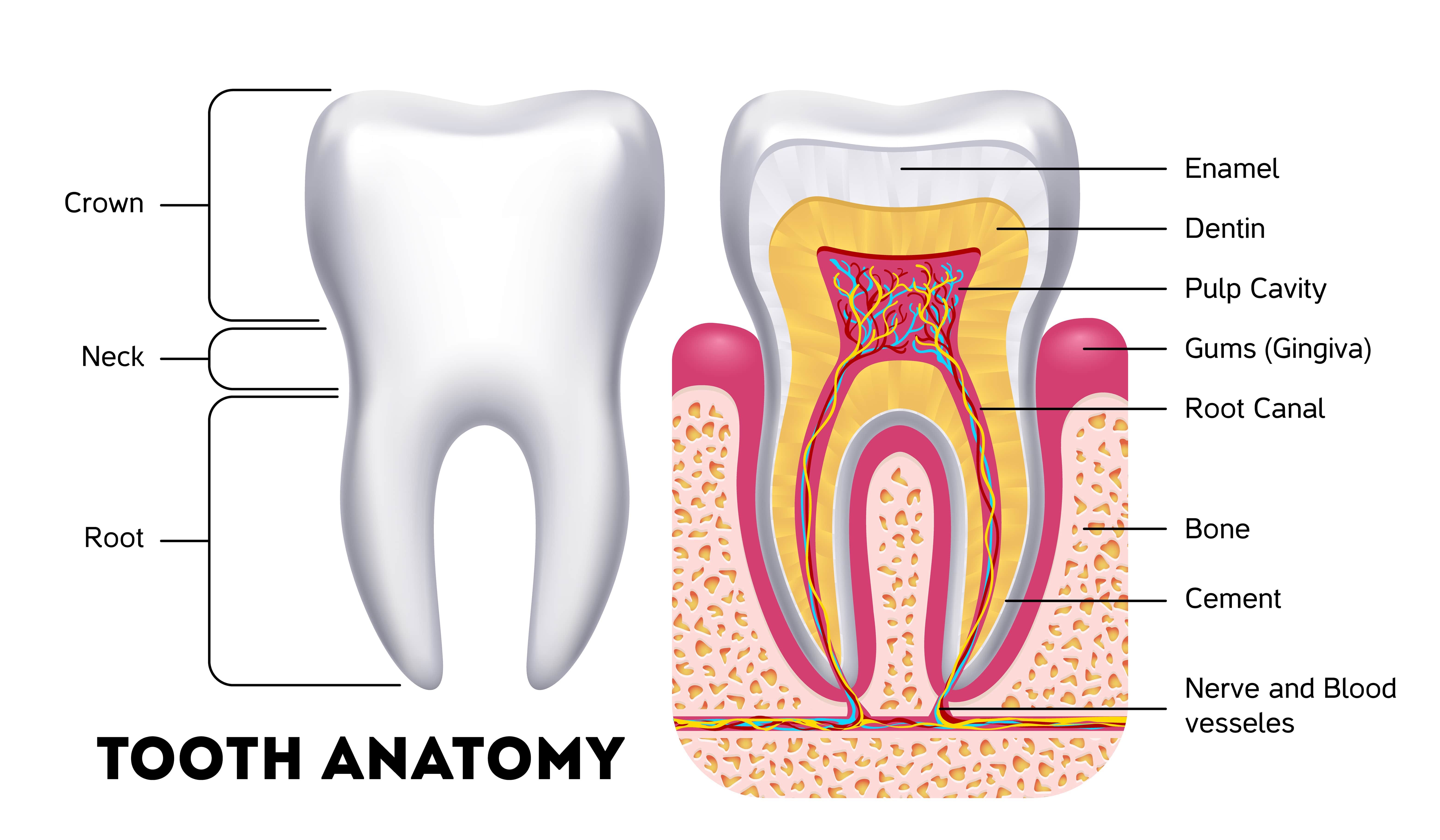
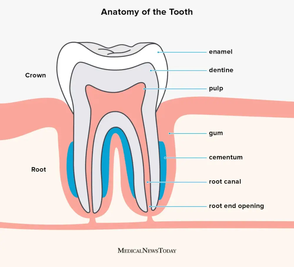
Learn about the four types of teeth, their parts, and how they work. Find out the common tooth conditions, symptoms, and tips for healthy teeth. Learn about the types, functions, structure and clinical aspects of the teeth with diagrams, videos and quizzes. The tooth is composed of enamel, dentin, pulp and cementum, and has a crown and a root.

Dental anatomy Biology Diagrams
Learn about the structure, function, and classification of human teeth with interactive 3D models. Explore the layers, cavities, and diseases of the tooth with detailed anatomy explorer.
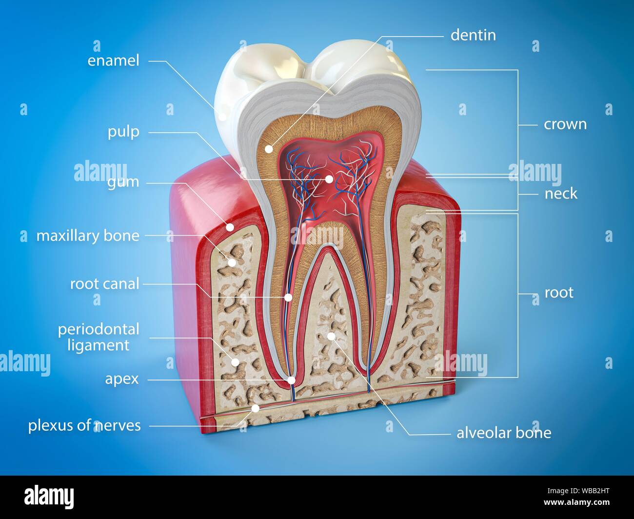
The teeth are aligned along the dental ridges and are surrounded by gingiva, forming the dental occlusion where the upper and lower teeth meet when the mouth is closed. Anatomy Each tooth is composed of multiple layers of tissues and is supported by surrounding structures like the periodontal ligament and gingiva. [1]
Anatomy atlas of the teeth Biology Diagrams
A comprehensive guide to teeth including types of teeth, tooth anatomy, tooth surface terminology and clinical relevance (e.g. tooth avulsion and enamel erosion). Tooth avulsion diagram. 9. Tooth anatomy. The tooth can be divided into two main components: the crown and the root (figure 7). The crown is the part of the tooth that is visible